")
")
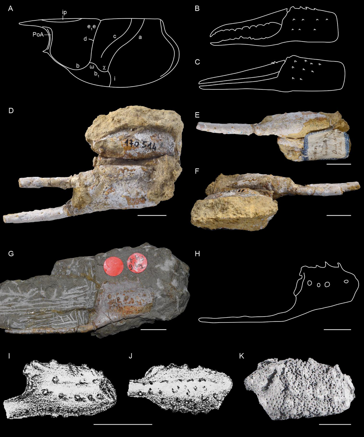
Fig. 14
Download original image
Morphology of the carapace and of the chela of the first pair of pereiopods of Stenodactylina Beurlen, 1928 and species from the Early Jurassic. A: typical carapace groove pattern of Stenodactylina; B: form I of chela of the first pair of pereiopods of Stenodactylina; C: form II of chela of the first pair of pereiopods of Stenodactylina; D–F: holotype FSL 170514 of Stenodactylina falsani (Dumortier, 1867) from the Sinemurian of Saint-Didier-au-Mont-d’Or (France): ventral view (D), external view (E), internal view (F); G–H: Holotype SMNS 7785 of Stenodactylina liasina Beurlen, 1928 from the Toarcian of Holzmaden (Germany): general view (G), line drawing (H); I–J: original figures of Étallon (1861: pl. 1, fig. 10) of the holotype of Stenodactylina spinosa (Étallon, 1861) n. comb. from the Toarcian of Les Nans (France): ventral view (I), outer view (J); K: original figure of Förster (1966: pl. 17, fig. 5) of a specimen of S. spinosa from the Toarcian of Perrigny (France). Scale bars: 1 cm. Abbreviations: a: branchiocardiac groove; b: antennal groove; b1: hepatic groove; c: postcervical groove; d: gastro-orbital groove; e1e: cervical groove; i: inferior groove; ip: intercalated plate; PoA: post-orbital area; χ: attachment site of adductor testis muscle; ω: attachement site of mandibular muscle. Photographs: L. Cazes (D–F), J. Devillez (G). Line drawings: J. Devillez.
Current usage metrics show cumulative count of Article Views (full-text article views including HTML views, PDF and ePub downloads, according to the available data) and Abstracts Views on Vision4Press platform.
Data correspond to usage on the plateform after 2015. The current usage metrics is available 48-96 hours after online publication and is updated daily on week days.
Initial download of the metrics may take a while.