")
")
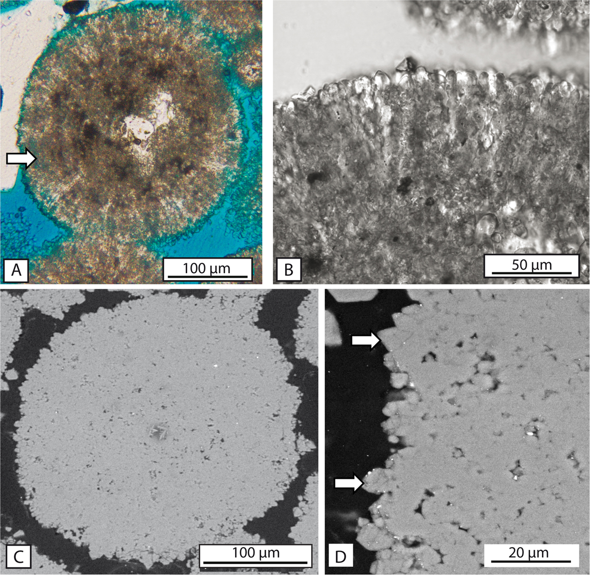
Fig. 5
Download original image
Microstructure of ooids. A) Thin-section microphotograph under polarized-light microscopy showing the variably preserved radiaxial microstructure within the cortex and the occurrence of micritized areas (arrows). The inner part of the ooid is pervasively micritized and the nucleus consists of a quartz grain. B) Detail of the radiaxial microstructure of ooid cortex under reflected-light microscopy. C) SEM photograph of a thin-sectionned micritized ooid showing the euhedral to subhedral micrite probably replacing the initially radiaxal microstructure and the presence of significant inter-crystalline microporosity. D) Close-up on the outer cortex of a micritized ooid under SEM showing microrhombic calcite overgrowths (arrow).
Current usage metrics show cumulative count of Article Views (full-text article views including HTML views, PDF and ePub downloads, according to the available data) and Abstracts Views on Vision4Press platform.
Data correspond to usage on the plateform after 2015. The current usage metrics is available 48-96 hours after online publication and is updated daily on week days.
Initial download of the metrics may take a while.