")
")
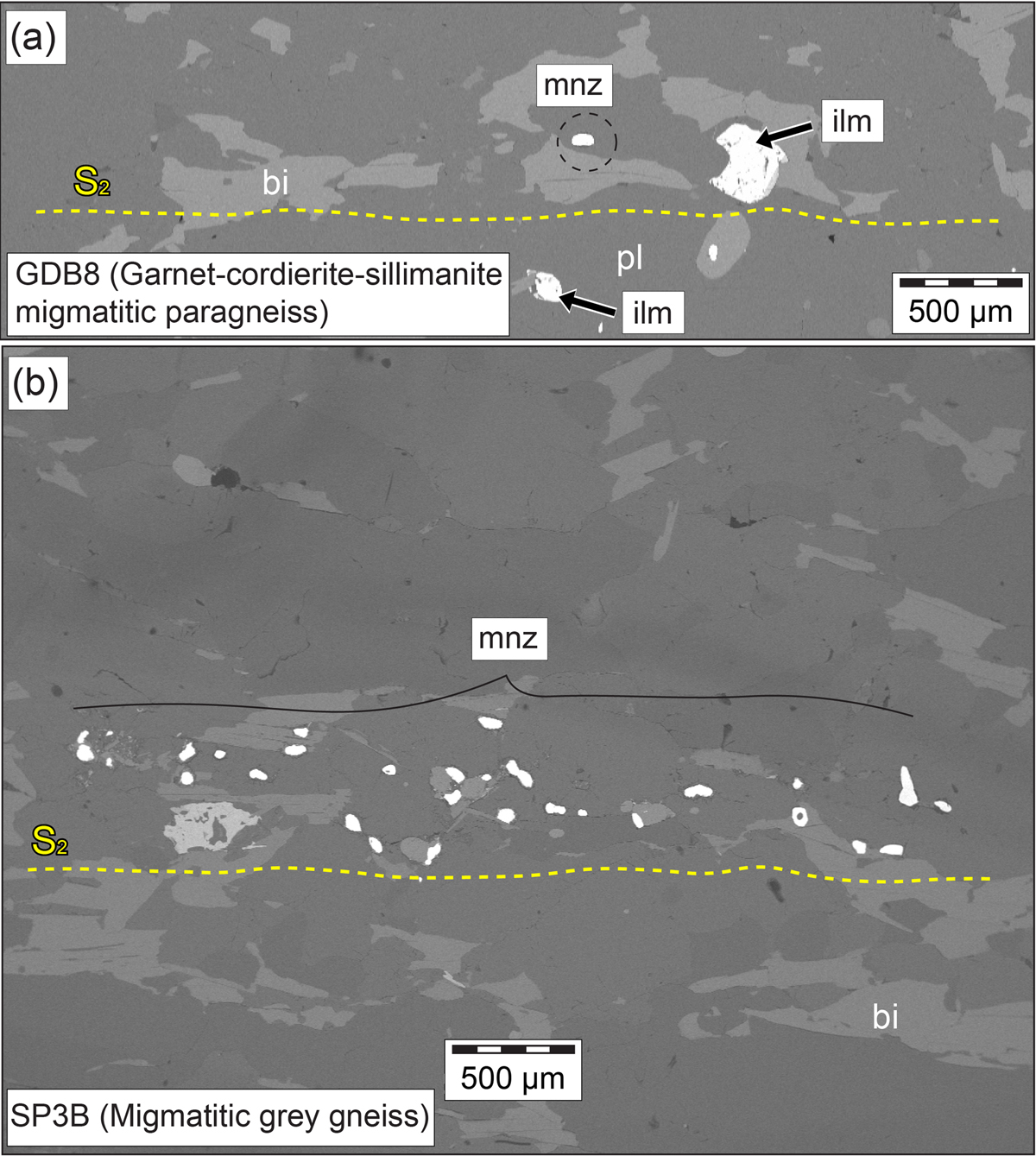
Fig. 11
Download original image
(a) Position of the monazite crystals (bright spots) in the thin section GDB8. (b) Alignment of monazite clusters parallel to S2 in migmatitic grey gneiss SP3B. (c–g) Migmatitic grey gneiss SP3B. (c) Back-scattered electron (BSE) images showing the position of the monazite crystals (bright spots) with respect to the garnet porphyroblasts. (d, e) Microprobe X-ray maps and (f, g) BSE images with analytical spots and associated 207Pb/206Pb dates of monazite grain marked in (a). LC: low concentration; HC: high concentration.
Current usage metrics show cumulative count of Article Views (full-text article views including HTML views, PDF and ePub downloads, according to the available data) and Abstracts Views on Vision4Press platform.
Data correspond to usage on the plateform after 2015. The current usage metrics is available 48-96 hours after online publication and is updated daily on week days.
Initial download of the metrics may take a while.